
Reviewed by Dr. Suprabath H. N., Consultant Fetal Medicine Specialist, 2026-11-12
This article is general information and does not replace advice from your obstetrician.
Growth scan (28 to 32 weeks), in short
A growth scan is an ultrasound, usually done at 28 to 32 weeks, that measures your baby’s head, abdomen and thigh bone to estimate weight and compare it with other babies of the same gestational age. It takes about 20 to 30 minutes. Most growth scans show normal growth, and the trend across scans matters more than any single number.
This fetal growth scan focuses on three things: your baby’s weight, the fluid around your baby and the blood flow to your baby. The anomaly scan covers anatomy in more detail. This guide explains each line of a growth scan report at 28 weeks or later, so the numbers make sense before your next maternity and pregnancy care visit.
Why the growth scan is done at 28 to 32 weeks
By the third trimester, growth rate becomes the most useful signal, because most structural development was already checked at the anomaly scan. The growth scan tracks whether your baby is gaining size at the expected pace for gestational age. It can pick up slow or excessive growth early enough to act on it.
The two scans answer different questions:
| Anomaly scan | Growth scan | |
|---|---|---|
| When | 18 to 22 weeks | 28 to 32 weeks |
| Main question | Is the anatomy developing as expected? | Is the baby growing at the expected pace? |
| Focus | Brain, spine, heart, kidneys, limbs, placenta | Estimated weight, fluid around the baby, blood flow to the baby (Doppler) |
In some pregnancies, an additional scan closer to 34 to 36 weeks lets the obstetrician confirm a trend. That is common and does not by itself signal a concern.
How to prepare for your growth scan
No fasting or full bladder is needed for a growth scan at 28 to 32 weeks. The most useful thing to bring is your earlier scan reports.
Do you need to fast?
No fasting is needed for a growth scan, which takes about 20 to 30 minutes. Eat and drink as you normally would on the day.
Do you need a full bladder?
A full bladder is not needed for a growth scan. From 28 weeks your baby is easily seen without any bladder preparation.
What to bring
Bring these to a third trimester growth scan:
- Your antenatal file
- All your previous scan reports: the early pregnancy (dating) scan, the NT scan and the anomaly scan report, plus any earlier growth scans, so the specialist can compare today’s measurements with your earlier ones
- Your obstetrician’s referral, if you are coming from outside
You do not need to bring fruit or juice. Unlike the anomaly scan, the growth scan does not depend on your baby’s position.
Who can come in with you
Under the PCPNDT Act, 1994, no attender is allowed inside the scanning room for any ultrasound scan. Your partner or family member can wait in reception.
How long it takes
Plan for 20 to 30 minutes for the scan, plus reception time. The growth scan does not depend on your baby’s position, so it is usually completed in one go.
What the growth scan measures
The growth scan combines several measurements into one picture. No single number defines your baby’s growth; the specialist reads the pattern across all of them.
| Measurement | What it tells the specialist |
|---|---|
| HC (head circumference) and BPD (biparietal diameter) | Head size |
| AC (abdominal circumference) | A key growth indicator, often the most sensitive to nutrition and placental function |
| FL (femur length) | Thigh-bone length, used with the others for the weight estimate |
| EFW (estimated fetal weight) | A calculated weight combining the measurements above, not a direct measurement |
| AFI (amniotic fluid index) | The volume of fluid around your baby, checked again at this stage |
| Placenta | Its position and general appearance |
| Doppler studies | Blood flow through the umbilical artery, the middle cerebral artery (MCA) in the baby’s brain and the ductus venosus, plus the CPR (cerebroplacental ratio), which compares brain and cord blood flow. Done at every growth scan at Nectar. |
| Presentation | Whether the baby is head-down, breech or transverse, which can still change before delivery |
How the numbers appear on your report
Head, abdomen and thigh-bone measurements are usually printed in millimetres, and EFW in grams. Many reports add a “gestational age equivalent” column, which converts each measurement into the week of pregnancy it would typically match, for example 29 weeks 2 days.
One measurement matching an earlier week than your dates is common and is a separate question from the weight percentile. Babies have different proportions, so the specialist looks at all the measurements together and at the EFW percentile. The percentile is calculated against a growth chart named on the report, and Nectar’s reports use the Hadlock chart.
If you are comparing reports from two centres, check which chart each one used. The same baby can sit at slightly different percentiles on different charts.
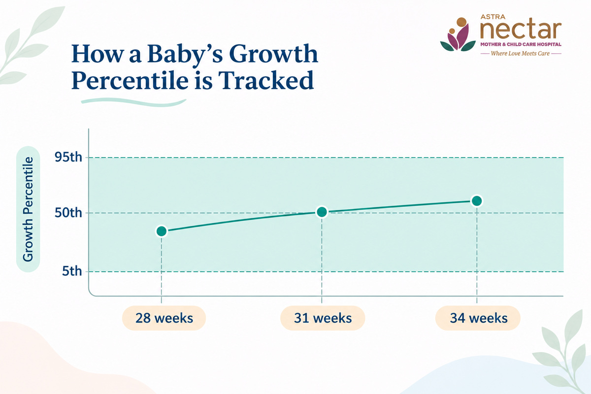
How to read a growth scan report at 28 weeks or 30 weeks: what percentile means
A percentile shows where your baby’s estimated weight sits compared with other babies at the same gestational age. The 50th percentile is the middle: half of babies at that age are smaller and half are larger.
A baby at the 30th percentile is heavier than about 30 of every 100 babies at that age. On the Hadlock chart used at Nectar, the 5th to 95th percentile range is considered normal. Any percentile inside that range can belong to a healthy baby.
| Term | What it means |
|---|---|
| Percentile | Your baby’s estimated weight ranked against babies of the same gestational age. 5th to 95th is the normal range on the Hadlock chart used at Nectar. |
| EFW (estimated fetal weight) | A calculated estimate with a built-in margin of error. Two scans a week apart can show slightly different numbers. |
| Trend | Whether the percentile holds steady across scans; see the example below. |
| AFI (amniotic fluid index) | Fluid volume in centimetres, read alongside growth because fluid and placental function are connected. |
| AGA (appropriate for gestational age) | The phrase most readers hope to see. It means growth is tracking within the expected range. |
A 2025 study in the International Journal of Gynecology & Obstetrics found that ultrasound weight estimates, on average, came in slightly below actual birth weight, by about 2.5 to 3.7 percent. For babies below the 10th percentile, the underestimate was larger: about 18 percent in the early third trimester and about 10 percent later on.
Estimates for babies below the 10th and above the 90th percentile were less accurate than for babies in between. An average error this small can still hide a wider spread for one baby, so a single EFW is best read as a range. For an individual baby, the estimate can differ from the eventual birth weight by about 10 percent in either direction.
Here is how the trend reads in practice:
| Scan | Baby A (illustration only) | Baby B (illustration only) |
|---|---|---|
| 28 weeks | 20th percentile | 60th percentile |
| 31 weeks | 22nd percentile | 35th percentile |
| 34 weeks | 19th percentile | 15th percentile |
| Reading (example values, not a real report) | A smaller baby tracking steadily: reassuring | Percentile falling across scans: prompts a closer look |
A single percentile is read together with your Doppler results, fluid levels, growth trend and pregnancy history, which help the specialist judge how much weight to give any single EFW. The same report-reading approach applies to our guide to the double marker test report.
What if your baby’s growth is outside the expected range?
Most growth scans in the third trimester confirm normal growth. When a measurement falls outside the expected range, the usual response is a closer look, often a repeat scan, before any change to your care plan.
A baby whose estimated weight falls below the normal range is described as small for gestational age (SGA). Many SGA babies are healthy and simply small. Fetal growth restriction (FGR) is the term used when a baby is not reaching their growth potential, which is identified when blood flow to the baby is compromised on Doppler or when the abdominal circumference measures below the 3rd percentile.
A baby whose estimated weight is above the 95th percentile is described as large for gestational age (LGA). Macrosomia refers to a birth weight above 4 kg. For a larger baby, the follow-up also includes a check of your blood sugar results, since diabetes in pregnancy is a common reason for faster growth.
The next steps usually run in this order:
- A follow-up scan after 2 weeks, for a baby whose growth is outside the normal range, whether small or large. If growth then looks normal, the specialist decides when the next follow-up is needed.
- Doppler studies of blood flow, which are part of every growth scan at Nectar and are repeated at the follow-up. Doppler shows how blood is moving through the umbilical cord, the baby’s brain and the ductus venosus, which helps the specialist judge whether the placenta is supporting growth.
- Closer monitoring or an earlier delivery plan, only if the trend or the Doppler results call for it.
At Nectar, obstetric care, fetal medicine and a Level 3 NICU are on the same campus, so any closer monitoring or delivery planning is coordinated in one place.
The growth scan exists to catch a trend early enough to plan, and a single number is rarely the whole story.
Book your growth scan at Nectar
Frequently asked questions
What is a growth scan and how is it different from the anomaly scan?
The anomaly scan, done at 18 to 22 weeks, checks your baby’s anatomy in detail: organs, spine, heart and limbs. The growth scan, typically done at 28 to 32 weeks, focuses on how well your baby is growing.
It uses head, abdomen and thigh-bone measurements to estimate weight and compare it with norms for your gestational age. Our fetal medicine team performs both.
Do I need to fast or drink water before a growth scan?
No. A third-trimester growth scan has no fasting or bladder-preparation requirement, so eat and drink normally on the day. The most useful thing to bring is your earlier scan reports, so the specialist can compare them with today’s measurements.
What does my baby’s percentile mean?
Percentile shows where your baby’s estimated weight sits compared with other babies at the same gestational age. The 5th to 95th percentile range is considered normal on the Hadlock chart used at Nectar. What matters more than one reading is whether your baby’s percentile stays consistent across scans.
The percentile depends on the growth chart named on your report, so two centres can print slightly different figures for the same baby.
Is a low percentile always a problem?
No. Babies naturally come in different sizes, and a baby who has tracked around the 15th percentile across several scans may simply be a smaller baby growing consistently well. The specialist watches for a percentile that drops significantly between scans, because a falling percentile is what prompts closer monitoring.
How accurate is the estimated fetal weight (EFW)?
For an individual baby, the estimate can differ from the eventual birth weight by about 10 percent in either direction, so it is best read as a range. It is less precise for very small or very large babies. Read across scans, it works as a trend indicator and gives a rough guide to birth weight.
Do I need more than one growth scan?
Often, yes. Many pregnancies have a growth scan around 28 to 32 weeks and a second closer to 34 to 36 weeks to confirm the trend. Your obstetrician will advise based on your pregnancy, and needing a second scan does not itself indicate a concern.
Will the growth scan tell me my baby’s sex?
No. Under India’s PCPNDT Act, 1994, disclosing the sex of the fetus at any stage of pregnancy is illegal at every ultrasound clinic in the country. Every scan at Nectar is conducted strictly for medical purposes, checking your baby’s growth and wellbeing, and our team is legally required not to disclose the baby’s sex at any scan.
What happens if my growth scan shows something outside the normal range?
Most growth scans confirm normal growth. Where a measurement is outside the expected range, the usual next step is a follow-up scan after 2 weeks. Doppler studies of blood flow are part of every growth scan at Nectar.
Because obstetric care, fetal medicine and a Level 3 NICU are on the same Nectar campus, any closer monitoring or delivery planning is coordinated in one place. A fetal medicine specialist can explain what your numbers mean.
Book your growth scan at Nectar
At our Kanakapura Road campus in South Bangalore, growth scans are performed and read by our in-house fetal medicine specialist, Dr. Suprabath H. N., with obstetric and NICU follow-up on the same campus if it is ever needed. To book, use the booking form below.
Already have a growth scan report from another centre? Call our helpline at +91 94800 20202 and our fetal medicine team can review it with you during your OPD appointment.